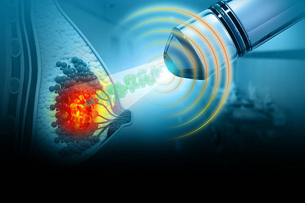
Understanding the Role of Early Imaging in Breast Cancer Detection
Breast cancer remains one of the most commonly diagnosed cancers among women worldwide, yet survival rates have improved dramatically over the past three decades. This progress stems largely from advances in early detection methods, particularly sophisticated imaging technologies that can identify abnormalities long before symptoms appear. While self-examination and clinical breast exams remain valuable, modern imaging techniques have transformed our ability to catch breast cancer at its most treatable stages.
The Foundation of Breast Screening
Mammography has served as the cornerstone of breast cancer screening for decades, using low-dose X-rays to create detailed images of breast tissue. Standard mammography can detect tumours as small as a few millimetres, often years before they become palpable. Women aged 40 and above are typically advised to undergo annual or biennial mammograms, though recommendations vary based on individual risk factors and family history.
Digital mammography has largely replaced traditional film-based methods, offering clearer images and the ability to manipulate contrast and zoom levels after the image is captured. This technology proves particularly beneficial for women with dense breast tissue, where traditional mammograms sometimes struggle to distinguish between healthy tissue and potential abnormalities.
Advanced Imaging Technologies
Tomosynthesis, often called 3D mammography, represents a significant leap forward in breast imaging. This technique captures multiple images of the breast from different angles, creating a three-dimensional reconstruction that allows radiologists to examine breast tissue layer by layer. Studies indicate that tomosynthesis can detect 20 to 65 percent more invasive breast cancers compared to standard 2D mammography, while simultaneously reducing false-positive results that lead to unnecessary callbacks and biopsies.
Ultrasound imaging serves as a crucial complementary tool, particularly for evaluating abnormalities found on mammograms or for screening women with dense breast tissue. Unlike mammography, ultrasound uses sound waves rather than radiation, making it safe for repeated use and valuable for distinguishing between solid masses and fluid-filled cysts. Automated breast ultrasound systems now allow for consistent, reproducible whole-breast screening that can be reviewed by multiple specialists.
Magnetic resonance imaging (MRI) of the breast offers the highest sensitivity for detecting breast cancer, though it comes with trade-offs in terms of cost and specificity. Breast MRI is typically reserved for high-risk women, such as those with BRCA1 or BRCA2 gene mutations, strong family histories, or prior chest radiation. The technique uses powerful magnets and radio waves to create detailed images of breast tissue, excelling at identifying cancers that other methods might miss.
When Screening Leads to Diagnosis
When screening reveals a suspicious finding, diagnostic imaging becomes essential for characterizing the abnormality and guiding treatment decisions. Diagnostic mammography involves additional views and magnification to examine specific areas of concern more closely. Radiologists may recommend spot compression views, which apply targeted pressure to spread out overlapping tissue and provide clearer visualization of a specific region.
For women in the United Arab Emirates seeking comprehensive breast health services, facilities offering diagnostic imaging in abu dhabi provide access to the full range of screening and diagnostic technologies. These services typically include mammography, ultrasound, MRI, and image-guided biopsy procedures, allowing for seamless transitions from screening to diagnosis and treatment planning when needed.
Image-guided biopsy techniques have eliminated much of the guesswork from breast tissue sampling. Stereotactic biopsy uses mammography to precisely locate an abnormality in three-dimensional space, while ultrasound-guided and MRI-guided biopsies serve similar purposes using their respective imaging modalities. These minimally invasive procedures allow pathologists to examine suspicious tissue without the need for surgical excision in most cases.
The Interplay Between Imaging and Lifestyle
While sophisticated imaging technologies detect breast cancer, prevention remains equally important. Research consistently demonstrates that lifestyle factors influence breast cancer risk, with diet, exercise, alcohol consumption, and body weight playing significant roles. Maintaining a healthy weight through balanced nutrition and regular physical activity may reduce breast cancer risk by up to 20 percent in postmenopausal women. For practical guidance on cancer-preventive habits, resources about keeping cancer away through diet and lifestyle modifications offer evidence-based strategies for reducing risk through daily choices.
The relationship between breast density and cancer risk illustrates why some women benefit from supplemental screening beyond standard mammography. Dense breast tissue not only makes mammography less sensitive but also represents an independent risk factor for breast cancer. Women with extremely dense breasts face a four to six times higher risk compared to those with mostly fatty tissue, making the choice of screening modality particularly important for this population.
Navigating Screening Recommendations
Screening recommendations vary among medical organizations, reflecting ongoing debates about the balance between benefits and potential harms. The American Cancer Society recommends annual mammograms starting at age 45, with the option to begin at 40, while other organizations suggest biennial screening starting at 50. These differences stem from varying assessments of false-positive rates, overdiagnosis, and the magnitude of mortality reduction achieved through screening.
Women with elevated risk factors should discuss personalized screening plans with their healthcare providers. Factors warranting enhanced surveillance include family history of breast or ovarian cancer, known genetic mutations, prior chest radiation therapy, history of atypical hyperplasia or lobular carcinoma in situ, and extremely dense breast tissue. High-risk women may benefit from earlier screening initiation, more frequent imaging, and the addition of breast MRI to their surveillance protocol.
Beyond Detection: Imaging in Treatment Planning
Once breast cancer is diagnosed, imaging continues to play a vital role in staging the disease and planning treatment. Accurate assessment of tumour size, location, and extent determines whether a woman is a candidate for breast-conserving surgery versus mastectomy. MRI proves particularly valuable for surgical planning, as it can reveal additional tumour foci that might otherwise be missed and help surgeons achieve clear margins while preserving as much healthy tissue as possible.
During treatment, imaging monitors response to therapy. Women receiving neoadjuvant chemotherapy, which aims to shrink tumours before surgery, undergo serial imaging to assess how well the cancer responds. This information helps oncologists determine whether to continue the current regimen or switch to alternative treatments. For comprehensive information about available resources and treatment approaches, breast cancer diagnosis and treatment guidance provides valuable context for patients and families navigating these decisions.
Making Informed Screening Decisions
The decision to undergo breast cancer screening involves weighing potential benefits against possible downsides. Benefits include reduced breast cancer mortality through early detection and treatment of aggressive cancers at more curable stages. Potential harms include false-positive results that cause anxiety and lead to additional testing, overdiagnosis of cancers that would never have caused symptoms during a woman's lifetime, and radiation exposure from repeated mammograms, though this risk remains very low.
Understanding your personal risk profile helps inform appropriate screening choices. Women at average risk may follow standard guidelines, while those with elevated risk require individualized approaches. Genetic counselling and testing for hereditary cancer syndromes should be considered by women with strong family histories, enabling them to make informed decisions about enhanced surveillance or risk-reducing interventions.
Breast cancer detection has evolved from crude examinations to sophisticated imaging technologies that save lives through early intervention. While no screening method is perfect, the combination of appropriate imaging techniques, risk-based screening strategies, and lifestyle modifications offers women powerful tools for breast health management. Regular communication with healthcare providers about individual risk factors and screening options ensures that each woman receives care tailored to her specific circumstances and needs.



Discussion (0)
No comments yet. Be the first to comment!
Leave a Comment
You need to login to post a comment.